Have you ever worried about a strange mole on your skin? Traditionally, checking it meant a small surgical cut and a long wait for lab results. But a new technology developed at Stanford is changing everything. Doctors may soon be able to scan your skin and get results in minutes—without ever breaking the surface.
Musumeci Online – The Podcast. It is perfect for driving, commuting, or waiting in line!
Seeing Beneath the Skin Without a Cut
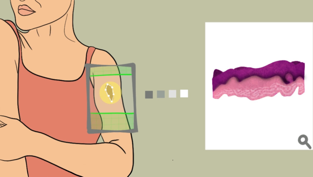
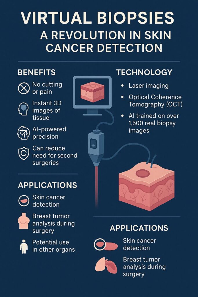
Thanks to advanced laser imaging and artificial intelligence (AI), researchers can now create detailed, 3D views of tissues inside the body. These “virtual biopsies” show the structure of skin and cells in such detail that they mimic the results of traditional lab slides used in cancer diagnosis.
Instead of taking a physical tissue sample and sending it to a pathologist, doctors can now scan the skin directly. The new method uses a refined form of optical coherence tomography (OCT)—a technique that reads how light bounces off tissue, kind of like how ultrasound uses sound to see inside the body. This method is already used by eye doctors, but the Stanford team made it powerful enough for use on skin and other tissues.
Why This Matters
Biopsies are routine but imperfect. Once tissue is cut and prepared on slides, it can’t be reused or looked at from different angles. Plus, patients usually have to wait days for results. Virtual biopsies change that.
- Instant results: No need to wait days for a lab report.
- No pain: It’s completely noninvasive—no cutting, no scarring.
- More information: The images show tiny cellular details that standard biopsies can miss.
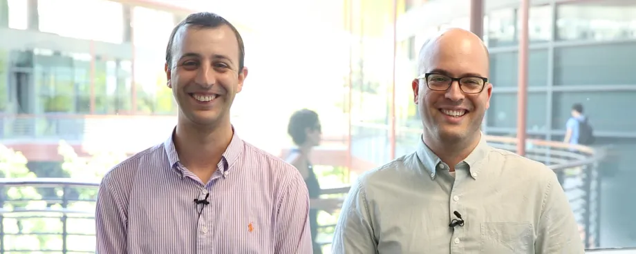
Imagine being in the dermatologist’s office and having a suspicious mole scanned and analyzed on the spot, right in front of you. That’s not science fiction—it’s becoming reality. In the picture: Dr. Adam de la Zerda (left) and Dr. Yonatan Winetraub (right)
Powered by AI
To make the new technology work in a real-world setting, the team taught an AI system to turn raw laser scans into images doctors are already familiar with. These images resemble the stained microscope slides (called H&E slides) that pathologists use every day to diagnose disease.
To train the AI, researchers scanned nearly 200 skin samples and matched them with actual lab-prepared slides. Over 1,500 image pairs were fed into the algorithm. The result? An AI that can now generate slide-like images from a simple scan, with a level of detail that doctors trust.
Helping Doctors Make Faster, Smarter Decisions
Three experienced dermatologists at Stanford tested the AI-generated images and confirmed they could see important cellular structures just as well as in traditional slides. Even better, because the images are digital, doctors can “slice” them in any direction—offering views never possible with real tissue samples.
This could be a game-changer, especially during surgery. For example, when surgeons remove a breast tumor, they usually send the tissue to a lab and wait to learn if all the cancer was removed. Sometimes, they miss a few cells and have to operate again. With virtual biopsies, doctors could get answers right away in the operating room, reducing the chance of repeat surgeries.
We didn’t just match the current gold standard—we improved it. – Dr. Adam de la Zerda
The Road Ahead
While more testing is needed before virtual biopsies become standard practice, the technology is progressing fast. It has the potential to not only transform skin cancer detection but also how we diagnose many other conditions.
As one of the lead researchers, Dr. Adam de la Zerda, put it: “We didn’t just match the current gold standard—we improved it.”
And that’s the real story: a future where diagnostics are faster, safer, and smarter—without the scalpel.
Leave a Reply